Dental practices use different types of equipment for oral examinations, diagnostic imaging, restorative procedures, periodontal treatment, root canal therapy, and instrument sterilization. Common dental equipment can be divided into four main categories: dental treatment units, imaging and diagnostic equipment, clinical treatment devices, and auxiliary and sterilization equipment.
Dental treatment equipment
Dental treatment units
A dental treatment unit is a basic piece of equipment in dental clinics and hospital dental departments. It is used for oral examinations, tooth cleaning, tooth preparation, and restorative procedures. A typical unit consists of a dental chair, instrument control system, operating light, air and water supply, and suction system.
Common accessories include high-speed and low-speed handpieces, a three-way syringe, a saliva ejector, and an instrument tray. The dental chair adjusts the patient's position, while the control system operates the handpieces and auxiliary instruments. The suction system removes saliva, cooling water, and debris generated during treatment.
The instrument configuration, control functions, and auxiliary features vary between dental treatment unit models.
Intraoral cameras
An intraoral camera typically consists of a miniature camera, illumination system, and image display. It captures images of tooth surfaces, gingiva, and other areas inside the mouth and displays enlarged views for examination.
Dentists can use these images to observe tooth cracks, dental caries, plaque, and gingival abnormalities. Real-time images can also help explain oral conditions to patients during consultations. An intraoral camera is an examination aid and does not replace clinical examination or necessary diagnostic imaging.
Dental imaging and diagnostic equipment
Panoramic dental X-ray machines
A panoramic dental X-ray machine uses panoramic tomography to produce a two-dimensional image of the upper and lower jaws, dental arches, and some surrounding anatomical structures.
It is commonly used for initial oral examinations, impacted tooth assessment, orthodontic evaluation, extraction planning, and preliminary dental implant assessment.
Panoramic images provide an overview of the oral structures, but they have limitations when detailed tooth root anatomy, complex bone structures, or three-dimensional relationships need to be examined. Some systems also offer imaging options for the temporomandibular joints, depending on the equipment configuration and acquisition method.
Cone beam computed tomography (CBCT) scanners
CBCT scanners use a cone-shaped X-ray beam to collect projection data from multiple angles. Computer reconstruction converts these data into three-dimensional volumetric images. Dentists can examine teeth, roots, alveolar bone, and jaw structures from different directions, reducing the limitations caused by anatomical overlap in two-dimensional images.
The field of view (FOV) and voxel size depend on the scanner model and acquisition protocol. Some equipment and scanning modes offer voxel sizes of 0.1 to 0.2 mm. These values should not be interpreted as a universal specification for actual spatial resolution or measurement accuracy.
CBCT is used for examinations with specific clinical indications, including preoperative implant assessment, complex root canal anatomy, impacted tooth localization, and oral and maxillofacial surgical planning.
Intraoral X-ray units
Intraoral X-ray units are used to produce localized two-dimensional dental images, including periapical and bitewing radiographs. During exposure, an image receptor is placed inside the patient's mouth, while the X-ray source remains outside and directs radiation toward the target teeth.
Periapical radiographs show the tooth crown, root, and surrounding periapical tissues. They help assess periapical lesions and root conditions. Bitewing radiographs are commonly used to detect interproximal caries and assess alveolar crest height and certain periodontal bone changes.
Unlike panoramic X-ray machines, intraoral units focus on specific teeth and provide detailed images of the selected area.
Clinical dental treatment equipment
Dental handpieces
Dental handpieces use pneumatic or electric drives to produce rotary motion. They are used for cutting tooth structure, preparing teeth, adjusting restorations, and polishing. Common types include high-speed and low-speed handpieces, which differ in rotational speed, transmission structure, and clinical applications.
High-speed handpieces commonly use air-turbine drives. Some models reach unloaded speeds of 300,000 to 400,000 rpm. They are mainly used for removing carious tissue, preparing teeth, and high-speed cutting during restorative procedures. Water spray is generally used for cooling to reduce heat generated during cutting.
Low-speed handpieces typically consist of a micromotor with a straight or contra-angle attachment. Common configurations operate at speeds of up to 40,000 rpm and are used for polishing, grinding, restoration adjustment, and selected tooth preparation procedures.
Actual handpiece speed depends on the pneumatic or electric drive, gear ratio, and specific model. The speed ranges of high-speed and low-speed handpieces should not be treated as universal product specifications.
Ultrasonic scalers
Ultrasonic scalers use piezoelectric or magnetostrictive transducers to generate high-frequency mechanical vibrations. The vibrating working tip removes dental calculus and plaque from tooth surfaces. These devices usually include a water supply system that cools the tip and flushes away debris during treatment.
Operating frequencies vary between models. Common equipment operates at approximately 25 to 36 kHz, depending on the instrument and working tip design.
Ultrasonic scalers are mainly used for supragingival scaling. Some systems and compatible tips can also be used for subgingival periodontal debridement. The appropriate tip and power setting depend on the patient's periodontal condition and the treatment requirements.
Dental light curing units
Dental light curing units emit light at selected wavelengths to activate photoinitiators in light-sensitive restorative materials. This initiates polymerization in materials such as composite resins, resin-based adhesives, and certain light-cured restorative products.
Common LED curing lights emit blue light within an approximate range of 430 to 480 nm. Common photoinitiators such as camphorquinone have a main absorption range of approximately 455 to 481 nm. Some curing units use multiple wavelengths to accommodate materials containing different photoinitiator systems.
A single exposure may take 20 to 40 seconds, but the required time depends on the restorative material instructions, light output intensity, distance between the curing tip and material, and material thickness. A fixed exposure time should not be applied to every restorative material.
Root canal treatment systems
Root canal treatment systems are used for working length measurement, canal preparation, and related endodontic procedures. Common equipment includes endodontic motors and electronic apex locators.
An endodontic motor drives nickel-titanium files in rotary or reciprocating motion. The operator can adjust rotational speed and torque according to the file system requirements. Some motors also provide torque control, automatic reverse, and reciprocating motion. Adjustable speed ranges vary between systems. Some units offer settings covering approximately 2 to 500 rpm, but the actual operating parameters should follow the equipment and file manufacturer's instructions.
An electronic apex locator estimates the working length of a root canal by measuring changes in electrical characteristics between the instrument inside the canal and an electrode placed on the oral mucosa. Modern devices commonly use alternating-current impedance measurements, multi-frequency impedance ratios, or other electrical measurement methods. Results can be affected by fluid inside the root canal, conductive substances, and anatomical conditions. Measurements should therefore be interpreted alongside clinical procedures and imaging findings. An accuracy specification of 0.5 mm or less should not be assumed to apply to every device under all conditions.
Dental laser equipment
Dental lasers use light at specific wavelengths to interact with teeth, gingiva, and other oral tissues. They are used for selected hard and soft tissue procedures, periodontal treatment, and certain adjunctive therapies.
Common types include Er:YAG lasers and semiconductor diode lasers. Er:YAG lasers operate at a wavelength of 2940 nm and interact with water and hydroxyapatite, making them suitable for selected dental hard tissue and soft tissue procedures.
Common semiconductor diode laser wavelengths include 810 to 980 nm. These systems are mainly used for gingival incision, gingival contouring, and selected periodontal and endodontic applications.
Laser systems differ in output power, pulse mode, energy settings, and clinical indications. Their use must be determined according to tissue type, treatment objectives, and the equipment manufacturer's instructions. The antimicrobial effect of laser treatment should not be considered equivalent to complete sterilization.
Auxiliary and sterilization equipment
Steam sterilizers
Steam sterilizers use saturated steam under controlled pressure, temperature, and exposure time to sterilize heat-resistant dental instruments. They are widely used in dental clinics and hospitals for instrument reprocessing.
Common sterilization programs include temperature settings of 121°C and 134°C. A 134°C cycle is commonly available for compatible dental instruments. However, the sterilization temperature, exposure time, and total cycle duration depend on the sterilizer model, instrument type, packaging method, and manufacturer's instructions. A universal cycle time of 15 to 30 minutes cannot be applied to every sterilization process.
Dental handpieces and other reusable heat-resistant instruments must be cleaned, lubricated, packaged, and sterilized according to their validated manufacturer's instructions. Mechanical, chemical, and biological monitoring methods are also used to verify sterilization performance.
Dental model fabrication equipment
Dental model fabrication equipment supports the design, production, and adjustment of dental restorations. Common machines include vacuum mixers, plaster trimmers, and dental laboratory polishing units.
A vacuum mixer combines gypsum or other model materials under reduced pressure to reduce air bubbles during mixing and improve model surface quality. A plaster trimmer is used to shape model edges and bases, while a dental laboratory polishing unit is used to finish restorations and related fabricated components.
These machines are mainly used in dental restoration and dental laboratory work. The equipment configuration depends on the workflow of the dental clinic or dental laboratory.
Custom precision machining for dental equipment components
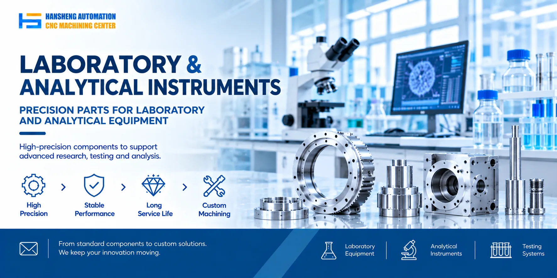
Dental equipment contains various mechanical structures and functional components. Examples include positioning brackets for imaging systems, connecting parts for dental treatment units, transmission components for treatment devices, and precision structural parts for model fabrication equipment. Each component has its own material, dimensional, structural, and assembly requirements.
For non-standard parts that require custom manufacturing, we provide precision machining based on customer-supplied engineering drawings to meet the specified manufacturing requirements.
Hansheng Automation also provides custom machining services for components used in other industries, including yacht structural parts, remote-control racing car components, tobacco packaging machinery parts, and filling machinery components. We manufacture non-standard parts according to customer drawings to support different equipment manufacturers with their component production and assembly requirements.